Advanced X-ray technology allows researchers to measure from synapse to whole brain level.
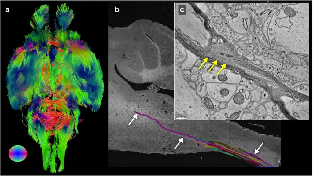
Researchers at the University of Chicago and the U.S. Department of Energy’s (DOE) Argonne National Laboratory have imaged a whole mouse brain across five orders of magnitude of resolution, which researchers say will better connect existing imaging approaches and uncover new details about the structure of the brain.
This research, published in NeuroImage, was accomplished in part using the resources of the Advanced Photon Source (APS), a DOE Office of Science User Facility. Scientists leveraged existing X-ray microscopy techniques to create a bridge between magnetic resonance imaging (MRI) scans and electron microscopy imaging, providing a pipeline for using multiple methods to image the same brain at different scales.
“Our lab is really interested in mapping brains at multiple scales to get an unbiased description of what brains look like,” said senior author Narayanan “Bobby” Kasthuri, assistant professor at UChicago and a scientist at Argonne. “When I joined the faculty here, one of the first things I learned was that Argonne has this extremely powerful X-ray microscope, and that it hadn’t been used for brain mapping yet, so we decided to try it out.”
Using X-ray tomography, researchers were able to image the entire mouse brain – roughly one cubic centimeter – at the resolution of a micron. It took roughly six hours to collect images, which is one of the fastest approaches for whole-brain imaging at this level of resolution.
“Why did we choose the mouse brain? Because it fits in the microscope,” Kasthuri said with a laugh. “But also, the mouse is the workhorse of neuroscience; they’re very useful for analyzing different experimental conditions in the brain.”
The X-ray data provides a bridge between MRI scans, which quickly image a whole brain at lower resolution, and electron microscopy, which reveals details of individual synapses, but generates an enormous amount of data.
“There have been a lot of imaging studies using MRI to look at the whole brain level and then try to validate those results using electron microscopy,” said first author Sean Foxley, assistant professor at UChicago. “It’s hard to say anything about the large volume of tissue you see with an MRI when you’re looking at an electron microscopy dataset, and the X-ray can bridge that gap.”
Researchers are using this technique to explore important questions in neuroscience, such as how Alzheimer’s disease develops.
For more information, visit this link.